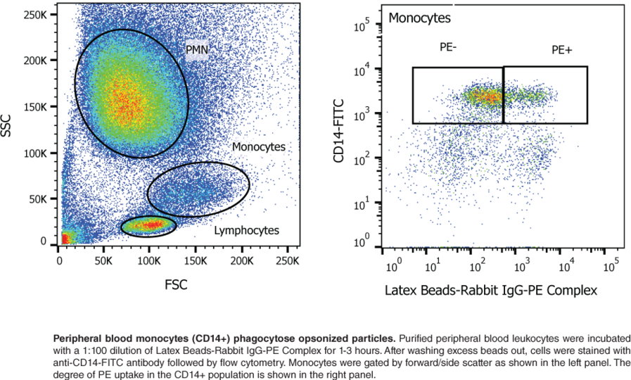
Phagocytosis Latex Beads
After 1 h of incubation uptake of the beads was stopped by incubating the cells on ice and the cells were analyzed by flow cytometry. Phagocytosisactivityinthe presenceof opsonized latex beads.
 Vav Is Not Required For Phagocytosis Of Latex Beads Or Bacteria A Scientific Diagram
From Phagocytosis latex beads

Readily phagocytose beads even without differentiation into macrophages.. The phagocytic assay consisted of two steps. Concentration-dependent uptake of beads. 1Department of Zoology Oregon State University Corvallis 97331-2914 USA.
However although phagocytosis of fluorescent latex beads by phagocytes have been studied for several decades 79 there has not been a reliable quantitative method to measure phagocytic ability of peripheral blood leukocytes for clinical application. The ability to purify latex bead-containing phagosomes has opened the door to allow comprehensive biochemical analyses and functional assays to study the molecular mechanisms governing phagosome function. The phagocytosis of latex beads by Acanthamoeba castellanii Neff involves a complex series of integrated events resulting in the selective inges-tion of one or more beads within a phagocytic vesicle.
Latex beads 1305 190 and 268 µ in diameter are ingested individually with each bead tightly surrounded by a membrane derived from the plasma membrane. Fryer SE1 Bayne CJ. Im hoping to carry out a phagocytosis assay with latex beads which I will coat with FITC-dextran.
25 µl of 200 times diluted latex beads either carboxylate-modified polystyrene fluorescent yellowgreen YG Sigma-Aldrich St. Caymans Phagocytosis Assay Kit IgG FITC employs latex beads coated with fluorescently-labeled rabbit IgG as a probe for the measurement of the phagocytic process in vitroThe engulfed fluorescent beads can be detected using a fluorescence microscope allowing kinetic studies of phagocytosis. Caymans Phagocytosis Assay Kit IgG FITC employs latex beads coated with fluorescently-labeled rabbit IgG as a probe for the measurement of the phagocytic process in vitro.
Many phagocytosis assays use IgG-coupled latex beads which are taken up after recognition by Fc receptors. Latex beads were labeled with either pHrodo red or pHrodo green and incubated with THP-1 cells at various concentrations. Phagocytosis of latex beads by the epithelial cells in the terminal region of the vas deferens of the cat.
L L Williams Department of Ophthalmology Ohio State University College of Medicine Columbus. Latex beads 0557 0264 0126 and 0088 micro in diameter are accumulated at the surface of the ameba and then phagocytosed with many beads tightly packed within one membrane-bounded vesicle. The aim is to measure bead uptake in LPS.
Recent advances in phagosome biology have been made possible largely by a model system that uses inert latex beads. After 1 h of incubation cells were washed and the mean fluorescence intensity was measured by flow cytometry. Phagocytosis of latex beads by Biomphalaria glabrata hemocytes is modulated in a strain-specific manner by adsorbed plasma components.
Technical advances in the assessment of phagocytosis have allowed rapid advan-. Best latex beads for macrophage phagocytosis assay. Latex beads 0557 0264 0126 and 0088 p in diameter are accumu- lated at the surface of the ameba and then phagocytosed with many beads tightly packed within one membrane-bounded vesicle.
Capacitance measurements made during phagocytosis of latex beads in macrophages led to the proposal that endomembranes were recruited at the cell surface for phagosome formation by a process referred to as focal exocytosis. The development of a quantitative assay allowed the study Weisman and Korn 1967 of a number of biochemical parameters of the proc-ess. Phagocytosis of latex beads is defective in cultured human retinal pigment epithelial cells with persistent rubella virus infection.
Latex beads labeled with pHrodo red A B or pHrodo green C D were added to THP-1 cells. Latex beads 1305 190 and 268 micro in diameter are ingested individually with each bead tightly surrounded by a membrane derived from the plasma membrane. Electron microscopic studies confirm and extend the conclusions derived previously from a quantitative biochemical study of the phagocytosis of polystyrene and polyvinyltoluene latex beads by Acanthamoeba 1.
In addition the flow cytometric readout provides the. These kits will allow you to analyze IgGmediated and Fc receptor-dependent phagocytosis. The engulfed fluorescent beads can be detected at the single-cell level.
Louis MO USA cat. SEM and TEM study. Murakami M Iwanaga S Nishida T Aiba T.
Phagocytosis of the pHrodo green-labeled beads was completely blocked by incubation on ice Figure 1D. Readily phagocytose beads even without differentiation into macrophages.
 Internalization Of Latex Beads In Phagosomes Phagocytic Assay In Human Scientific Diagram
 Phagocytosis Of Cypher5e Labeled Particles A Phagocytosis Of 3 Mm Scientific Diagram
 Color Phagocytic Assay Ingestion Of Latex Beads 0 45 µm By Scientific Diagram
 Phagocytosis Assay Kit Igg Pe Cayman Chemical
 Figure 2 From Quantifying Fitc Labeled Latex Beads Opsonized With Photoreceptor Outer Segment Fragments An Easy And Inexpensive Method Of Investigating Phagocytosis In Retinal Pigment Epithelium Cells Semantic Scholar
 Pma Activated Thp 1 Cells Allow Investigation Of Phagocytosis Of Scientific Diagram
 Phagocytosis Assay Of Engrafted Cells Four Hours After Latex Beads Scientific Diagram
 Phagocytosis Of Latex Beads By Intervertebral Disc And Other Cell Scientific Diagram
 Opn Enhances Phagocytosis Of Latex Beads Via The X Integrin Receptor Scientific Diagram
 Trpv4 Mediates Lps Stimulated Macrophage Phagocytosis Of Igg Coated Scientific Diagram
 Trpv4 Mediates Lpsstimulated Macrophage Phagocytosis Of Igg Coated Scientific Diagram
 Phagocytosis By Cd4 1 And Cd4 1 2 Myeloid Cells With Fluorescent Scientific Diagram
 Phagocytic Activity Ingestion Of Fluorescent Carboxylated Latex Beads Scientific Diagram
 In Vivo Phagocytosis Of Fluorescent Latex Beads And Fluorescent E Coli Scientific Diagram
 Microglial Phagocytosis Assay Bio Protocol
 Phagocytosis Of Latex Beads Notes Counting Of Macrophage Phagocytic Scientific Diagram
 Analysis Of Phagocytosis Inhibition Using Cypher5e Latex Beads A Scientific Diagram
Latex Beads Covalently Linked To Cps Inhibit The Phagocytic Capacity Of Scientific Diagram
 Protonex Red 600 Latex Bead Conjugate Aat Bioquest
 Phagocytosis Of Latex Beads By Luteal Macrophages A The Cell Scientific Diagram
 Phagocytosis Of Latex Beads By Luteal Macrophages A The Cell Scientific Diagram
 Figure 2 From Fusion Fission And Secretion During Phagocytosis Semantic Scholar
 Latex Beads Phagocytosis By Bm Mscs And Rpe Cells The First Picture In Scientific Diagram