Microfluidic Magnetic Bead Assay For Cell Detection
We present a novel cell detection device based on a magnetic bead cell assay and microfluidic Coulter counting technology. We designed an assay that fluorogenically detects ligation products following a simple magnetic separation through a microfluidic channel.

See more ideas about Microfluidic magnetic bead assay for cell detection

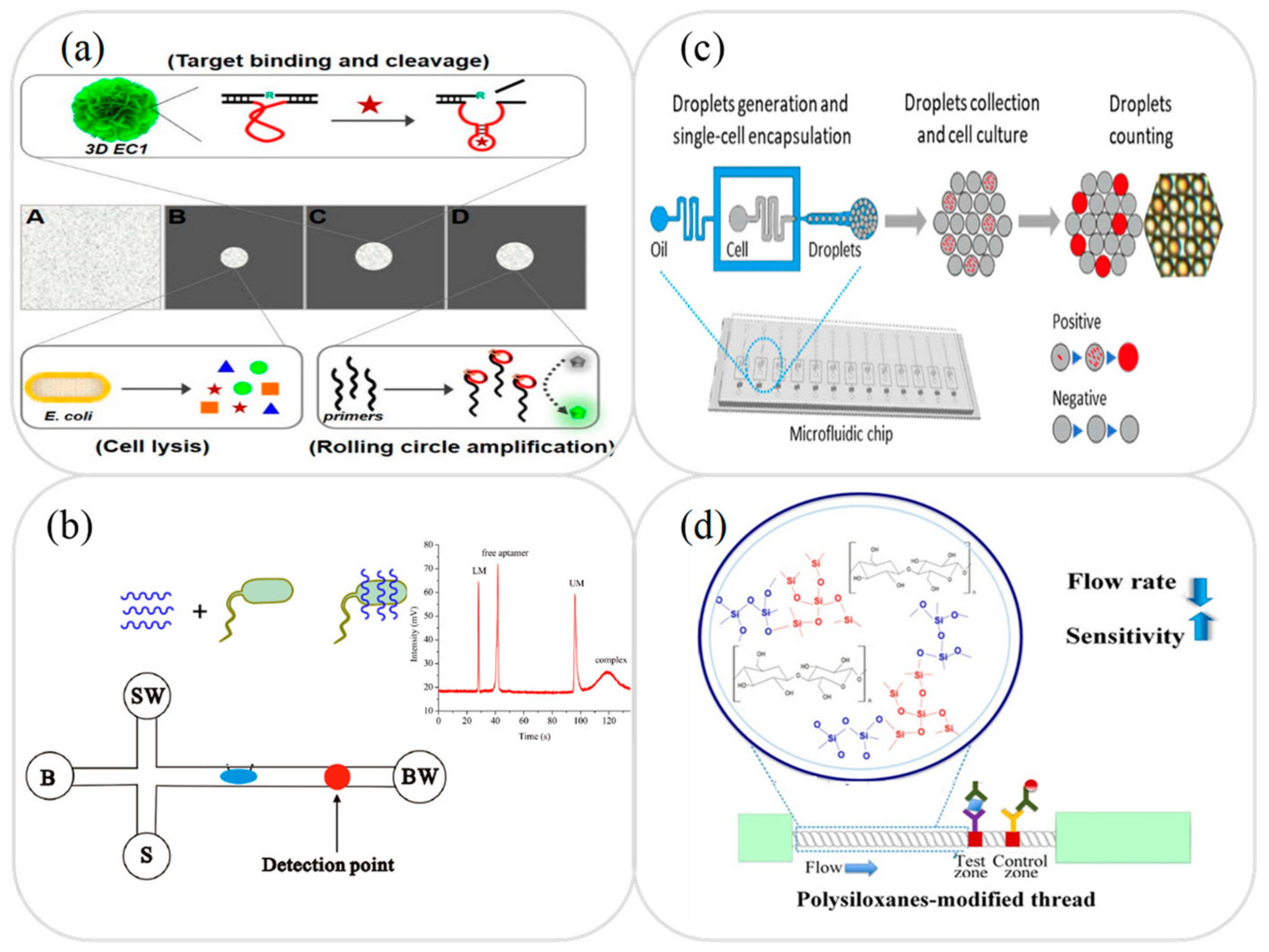
Here a microfluidic chip MC-based multianalysis method coupled with magnetic encoded aptamer probes was used for simultaneous detection of kanamycin 17β-estradiol and lead ion Pb 2.. During recent years magnetic beads have been used frequently in immunoassays either as mobile substrates on which the target antigen is captured as detection labels or simultaneously as substrates and labels. The device can detect specific target cells ratios as well as cells size distribution and concentrations. A lab-on-a-chip integrated microfluidic cell has been developed for magnetic biosensing which is comprised of anisotropic magnetoresistance AMR sensors optimized for the detection of single magnetic beads and electrodes to manipulate and sort the beads integrated into a microfluidic.
Create a account to . Using this innovative strategy the magnetic bead MB-based encoded probes labeled with aptamer hybrid chains were first used to selectively capture multiple targets followed by generating. The device can detect specific target cells ratios as well as cells size distribution and concentrations.
The device consists of two identical micro Coulter counters separated by a fluid chamber where an external magnetic field is applied. 1Department of Mechanical Engineering University of Akron Akron Ohio United States of America. A case for microfluidic magnetic bead-based assays.
The capture efficiency can reach up to 90 at 3mlh coated with immune beads and at 15 mlh without beads. In situ single cell detection via microfluidic magnetic bead assay. The device cannot only accuratelymeasure cells size distribution and concentration but also detect specific targetcells.
N3 B All MCF-7 cells were coated with magnet beads in bright field. Lab Chip 13 47114739 2013. 1016 Their simple separation and conjugation with a wide array of biomolecules has led to their integration in various detection methods.
Request Target Cell Detection via Microfluidic Magnetic Beads Assay We present a novel cell detection device based on a magnetic bead cell assay and microfluidic Coulter counting technology. With the compact size and simple operation steps this method is capable of single cell detection in a continuous flow and enables cell reuse for. Liu F1 Kc P2 Zhang G2 Zhe J1.
Rapid microfluidic separation of magnetic beads through dielectrophoresis and magnetophoresis. We present a novel cell detection device based on a magnetic bead cell assay and microfluidic Coulter counting technology. We demonstrated the feasibility of parallel detectiontrapping of different beads on the same chip.
The device consists of two identical micro Coulter cou. Microfluidics has been widely applied in genomics proteomics and cell cytometry to enable rapid and automated assays. To overcome the above limitation here we report a microfluidic magnet bead assay that can not only accurately identify the single cell in situ but also measure the sizes of individual cells and accurately count the cells within a mixed population.
We present a novel cell detection device based on a magnetic bead cell assay and microfluidic Coulter counting technology. First we introduce a simple and fast method for creating protein micropatterns both on a bare substrate and in-situ inside a microfluidic channel in a matter of minutes through electrostatic self-assembly of pre-functionalized magnetic beads. 100 nM of synthetic HIV-1 K103N minority mutant templates were successfully detected in 30 min.
C An MCF-7 cell was surrounded by immune magnetic beads. A new gravity-driven microfluidic-based electrochemical assay coupled to magnetic beads for nucleic acid detection. In this thesis we demonstrate microfluidic bio-assays based on novel methods for generating protein-patterns and on sensitive detection of the bio-targets.
Magnetic Particle Plug-Based Assays for. CAS Article Scholar. This separation approach offers the performance of multiplex analysis in labonachip devices.
We present a novel cell detection device based on a magnetic bead cell assay and microfluidic Coulter counting technology. For example magnetic microspheres have been used in thermotherapy for cancer cells immunoassays cell separation DNA hybridization protein and peptide separation and pathogen detection. Full Package.
To overcome the above limitation here we report a microfluidic magnet bead assay that can not only accurately identify the single cell in situ but also measure the sizes of individual. Microbeads have a high density of surface functional groups making them easy to modify and thereby making it possible to use higher concentrations of samples. The device cannot only accurately measure cells size distribution and concentration but also detect specific target cells.
2Department of Biomedical Engineering University of Akron Akron Ohio United States of America. with with Facebook. In situ single cell detection via microfluidic magnetic bead assay - Fig 2 By Fan Liu 27437 Pawan KC 1444732 Ge Zhang 112487 and Jiang Zhe 690388 Cite.
We review the use of magnetic micro- and nanoparticles magnetic beads in microfluidic systems for ultrasensitive protein detection. Bead binding is an important step in the magnetic-based isolation. Here a microfluidic chip MC-based multianalysis method coupled with magnetic encoded aptamer probes was used for simultaneous detection of kanamycin 17β-estradiol and lead ion Pb 2.
Droplet Microfluidic Workflow For Single Cell Highthroughput Screening Scientific Diagram
 Forces Acting On The Magnetic Beads A Scheme Of Microfluidic Scientific Diagram
 Design Of The Microfluidic Cell Separation Device The Device Consists Scientific Diagram
 Recent Advances In Microfluidic Platforms For Single Cell Analysis In Cancer Biology Diagnosis And Therapy Sciencedirect
 Structure And Working Principle Of The Microfluidic Device For The Scientific Diagram
Plos One In Situ Single Cell Detection Via Microfluidic Magnetic Bead Assay
 Microfluidic Devices For Exosome And Cell Dna Research A Scientific Diagram
 Microfluidic Chip Design And Functionality For Single Cell Protein Scientific Diagram
 A New Sensitive And Rapid Assaying Technique Biomedical Engineering Magnetic Beads Development
 Automatic Detecting And Counting Magnetic Beads Labeled Target Cells From A Suspension In A Microfluidic Chip Song these cool Electrophoresis Wiley Library
 Microfluidics Based Immunomagnetic Devices For Detection Of Scientific Diagram
 Micromachines Full Text Recent Advances In Microfluidic Devices For Contamination Detection And Quality Inspection Of Milk
 Design Of The Microfluidic Cell Separation Device The Device Consists Scientific Diagram
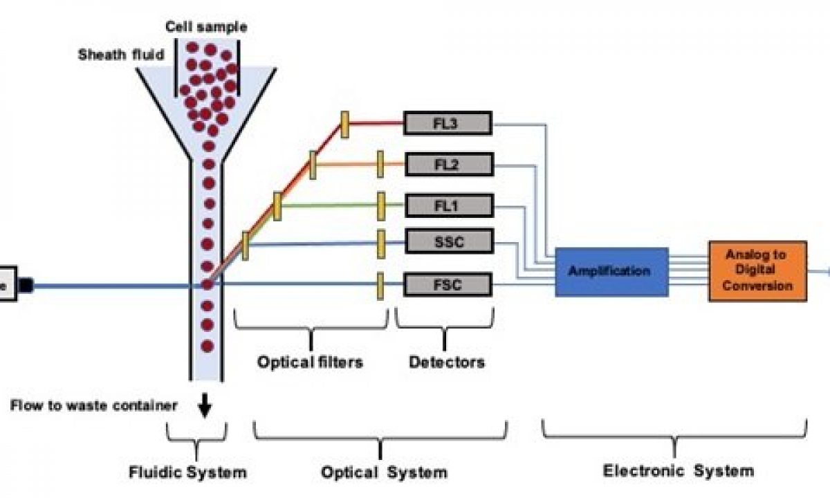 Microfluidic Flow Cytometry Principles And Commercial Review Ufluidix
Plos One In Situ Single Cell Detection Via Microfluidic Magnetic Bead Assay
 The Experimental Procedure Performed On The Integrated Microfluidic Scientific Diagram
 Microfluidic Cell Lysis Systems Using Different Energy Sources To Scientific Diagram
 Printed Electrode Chip And Microfluidic Flow Cell Design A Schematic Scientific Diagram
 Microfluidic Chips For Cell Trapping A Schematic Of A Single Ctc Scientific Diagram
3
 Schematic Illustration Of The Microfluidic Chip For Hsc Detection Scientific Diagram
 Microfluidic Communicating Vessel µcove Chip For Rapid Magnetic Scientific Diagram
 Digital Microfluidic Device For Cell Culture And Impedance Sensing A Scientific Diagram
 Microfluidic Devices For Exosome And Cell Dna Research A Scientific Diagram