Streptavidin Beads Elution
Harsh elution conditions or direct on-bead digestion promotes the release of streptavidin from the matrix thus generating a massive contamination of unwanted streptavidin peptides upon protease digestion. The use of this purification approach can be relatively costly.
 Overview Of The Streptavidin Pulldown Procedure Major Steps For The Scientific Diagram
Unique idea Streptavidin beads elution

MRNA Isolation using Streptavidin Magnetic Beads NEB S1420 and NEB S1421 For use with Streptavidin Magnetic Beads and Hydrophilic Streptavidin Magnetic Beads Introduction.. The unique DNA haploid binding to corresponding probe was isolated after washes and elution. The elution strat- egy was tested on different types and brands of streptavidin-coated magnetic beads. The elution of peptides from the beads is achieved by first mixing the beads with the MALDI matrix solution and removing after a few minutes the beads with a magnet.
The yield of polyA RNA will vary with the type of tissue or cells used. Non-denatured and active proteins from the cytosolic fraction of mesothelioma cells are used as the source of proteins. Then the matrix solution containing the biotinylated peptide is directly mass analyzed by.
The RNA-desthiobiotin is immobilized through interaction with streptavidin on magnetic beads which are used to pull down proteins that specifically interact with the RNA of interest. Regeneration efficiency using streptavidin beads from different sources. However as streptavidin binds biotin tightly K d 10 14 M the streptavidin matrix essentially can be used only once.
To test whether excess biotin or heat is required for efficient elution of biotinylated proteins from streptavidin conjugated beads the bound biotinylated proteins were eluted in 0 mM or 25 mM of biotin or 4 sample buffer and either left at room temperature or at 95 C for 5. Harsh elution conditions or direct on-bead digestion promotes the release of strep-tavidin from the matrix thus generating a massive contamination of unwanted streptavidin peptides upon protease digestion. Elution conditions B for the elution of the proteins remaining on the beads after the application of conditions A.
Interestingly the concentrations of SDS and IGEPAL-CA630 during the incubation with streptavidin conjugated beads were the key to effective elution of biotinylated. Here we use biotinylated bovine serum albumin as a working model to demonstrate a simple and rapid method for biotin-tagged protein purification under non-denaturing conditions. Streptavidin resins represents a critical drawback.
Streptavidin beads were categorized according to 1 the rate at which they form a tight pellet when placed near a magnet 2 whether cell pellets smear when aspirating supernatant and 3 how easily bead pellets can be resuspended in binding or elution buffer as this defines how the beads should be handled Table 2. Four con- secutive captures and releases of DNA fragments were performed using binding in 1. S4 SDS-PAGE of the BSA-linker 6 S8 or S9 conjugate eluted from the streptavidin beads.
Sample buffer 100 C 5 min. However if the Dynabeads Streptavidin have been used in applications such as isolation of DNA binding proteins and release of proteins from DNA is done by gentle methods like using high salt buffers that do not interfere with biotin-streptavidin interaction the beads with immobilized probe may be reused. Extraction of biotinylated peptides by streptavidin magnetic beads has been directly coupled to the MALDI-TOF mass analysis.
Biotinylated antibodies eg Goat Anti-Human Biotinylated IgG Cat V7830 or antigens. Single-stranded DNA ssDNA aptamer preparations eluted from biotinstreptavidin beads using NaOH during systematic evolution of ligands by exponential enrichment SELEX commonly suffer from streptavidin and double-stranded DNA dsDNA contamination. Biotin added to the elution buffer acts as an effective competitor to elute the bound SBP-tagged proteins off.
To effectively release biotinylated proteins bound to streptavidin conjugated beads we designed a series of experiments to determine optimal binding and elution conditions. To effectively release biotinylated proteins bound to streptavidin conjugated beads we designed a series of experiments to determine optimal binding and elution conditions. FAX 49 6221-764620 Email.
If SDS-PAGE buffer is selected for elution the eluate will contain streptavidin monomers and dimers and biotinylated antibody combined with target antigen. Low-pH elution buffers are. For the isolation of mRNA from 100 µg of total RNA or 5 x 10 6 cells.
If a low-pH elution buffer is selected for elution streptavidin might leach from the beads. As a result chromatography and subsequent mass spectrometry are readily. Streptavidin Beads ideal for use with complex biological samples.
100 μM 8 50 mM MPAA 40 mM TCEP 01 SDS 400 mM Na phosphate buffer pH 74 37 C 24 h. Fluorescence imaging λex 460 nm. The probe was mixed and extended with corresponding genomic DNA and incubated with streptavidin magnetic beads which could form a streptavidin magnetic beads-biotin-probe DNA complex.
Based on an extensive literature review most protocols describe elution with NaOH concentrations 100 mM. MRNA Isolation using Streptavidin Magnetic Beads. High Capacity Magne Streptavidin Beads can be used for a variety of applications including pharmacokinetics studies of therapeutic antibodies.
Interestingly the concentrations of SDS and IGEPAL-CA630 during the incubation with streptavidin conjugated beads were the key to effective elution of biotinylated proteins using excess biotin and heating. Infopolysciencesde TECHNICAL DATA SHEET 753 Streptavidin-Coated Microspheres Binding Biotinylated DNA Description The streptavidin-biotin bond is one Capture of ds Biotinylated DNA Elution of ssDNA 1. Aliquot 100µl streptavidin-coated microspheres into a microfuge tube.
The use of avidin or streptavidin in the purification of biotinylated proteins has been highly restricted due to the harsh and denaturing elution conditions. MRNA Isolation using Streptavidin Magnetic Beads NEB S1420 and NEB S1421 For use with Streptavidin Magnetic Beads and Hydrophilic Streptavidin Magnetic Beads Introduction.
 Sequence Specific Dna Capture With Streptavidin Nanobeads Nvigen
 Characterization Of A Dual Biotin Tag For Improved Single Stranded Dna Production Analytical Methods Rsc Publishing Doi 10 1039 C3ay41899e
A Binding Biotinylated Gata 1 To Streptavidin Beads Specifically Scientific Diagram
 How To Isolate Proteins And Peptides With Streptavidin Magnetic Beads
Onlinelibrary Wiley Doi 10 1002 Elps 200410070
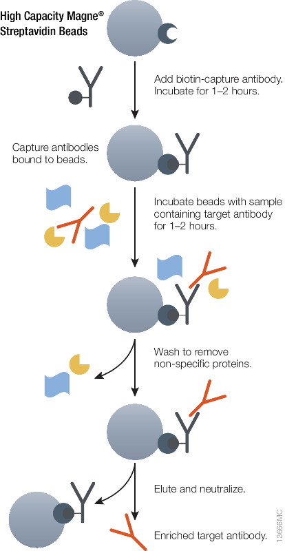 Optimizing Antibody Enrichment For Pharmacokinetic Assays Promega Connections
 High Capacity Magne Streptavidin Beads And Goat Anti Human Biotinylated Igg
 Exocap Streptavidin Kit Mblライフサイエンス
 Capturing Biotinylated Proteins Using Magnetic Streptavidin Beads Scientific Diagram
 Schematic Representation Of The Sbp Based Pull Down Technique For The Scientific Diagram
 Capturing Biotinylated Proteins Using Magnetic Streptavidin Beads Scientific Diagram
 A Simple Elution Strategy For Biotinylated Proteins Bound To Streptavidin Conjugated Beads Using Excess Biotin And Heat Abstract Europe Pmc
 Streptavidin Magnetic Beads Advanced Biochemicals
.jpg) Streptavidin Coated Magnetic Beads Biocompare Kit Reagent Review
Streptavidin Coated Magnetic Beads Biocompare Kit Reagent Review
 Fg Beads Drug Elution
Streptavidin Bead Pulldown Assay To Determine Protein
 µmacs And Multimacs Streptavidin Kits Biotinylated Molecule Isolation Nucleic Acid And Protein Isolation And Analysis Macs Molecular Analysis Products Miltenyi Biotec Usa
 Double Elution With Formic Acid Combined With Urea Improved The Elution Scientific Diagram
 Biotin Labeling Streptavidin Purification And Elution Of Proteins Scientific Diagram
 Streptavidin Mag Beads
1
 Biotin Streptavidin Affinity Pulldown Assay Of Rcz12 20 With Whole Cell Scientific Diagram
 Streptavidin An Overview Sciencedirect Topics
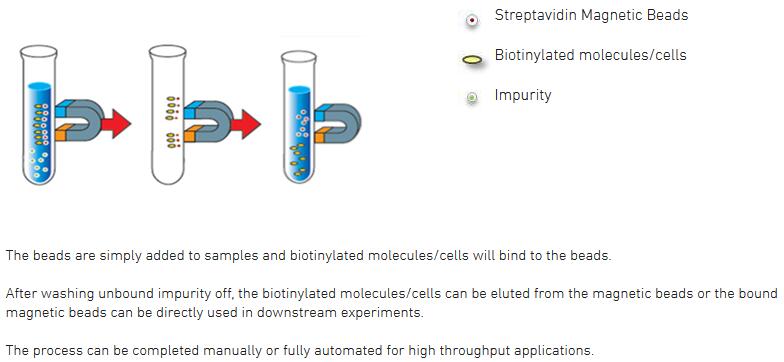
 Purification Or Removal Of Biotin And Biotinylated Biomolecules With Magnetic Beads