Human Igg Coated Beads
The coated polystyrene particles from Spherotech can be used in a variety of applications. For detection of IgG binding beads were incubated with 1 µgmL goat anti-human kappa-AlexaFluor647 Southern.
 Cbd Coated Beads Do Not Lead To Agglutination Of Cell Bead Mixtures Scientific Diagram
Human igg coated beads

The magnetic beads are incubated with a sample containing antibodies and then held by a magnet next to the tube.. Beads coated with ligands IgG TDM or β-glucan to measure acidification and proteolysis activities were prepared as previously described Yates and Russell 2008. AlphaLISA Acceptor beads conjugated to anti-human IgG antibody. Magnetic Dynabeads M-270 Streptavidin Invitrogen 6 to 7 10 5 2 05 mM CaCl 2 005 volvol Tween 05 HSA dependent on the buffer used in the subsequent incubation step.
Protein G exhibits high affinity for subclasses of IgG from many species including human rabbit mouse rat and sheep 1. The biology and function of IgG has been extensively reviewed in the literature. They also react with light chains of other mouse immunoglobulin classes like IgM IgA or IgE.
Protein G exhibits high affinity for subclasses of IgG from many species including human rabbit mouse rat and sheep 1. The antibody conjugated to these beads is Fc-specific and is a. Ad Unlabeled or conjugated.
The high capacity of the beads enables enrichment of antibodies over a wide concentration range using small amount of beads. AF ATTO Biotin HRP FITC PerCP RPETRITC etc. Anti-Mouse IgG MicroBeads are conjugated with goat Fab.
Ad Unlabeled or conjugated. It consists of a biotinylated Nanobody that specifically binds to human IgG antibodies with high affinity and in a site-directed manner. The conjugated antibody targets the Fc region of human IgG.
Tram et al 2019. Protein G Magnetic Beads are an affinity matrix for the small-scale isolation and purification of immunoglobulins. They are utilized in the magnetic separation and isolation of human antibodies from serum or human antibody-labeled components.
A truncated form of recombinant Protein G is covalently coupled to a nonporous superparamagnetic particle. These beads can be used to capture human IgG antibodies. Anti-Human IgG Magnetic Beads are densely coated with Goat anti-Human IgG.
The BD CBA Human Total IgG Flex Set is a bead-based immunoassay capable of measuring human total immunoglobulin G IgG in serum and plasma samples. On the other hand the 07-09 μm Goat anti-Mouse IgG coated polystyrene particles are prepared using passive adsorption. Similarly the avidin and biotin coated particles are also prepared by covalent coupling.
Human IgG at increasing concentrations was coated onto beads and detected with an alkaline phosphatase-conjugated anti-human IgG. After the enrichment of B cells with the CD19 MultiSort Kit IgG-expressing memory B cells are specifically labeled and isolated with Anti-IgG MicroBeads. Anti-human IgG Fc specific AlphaLISA Acceptor Beads 250 µg.
For more information on bead-based immunoassays refer to. The results show that within a certain range of concentrations of the added ligand there is a direct correlation to the amount that actually adsorbs to the magnetic beads. Enrichment or depletion of memory B cells that express surface IgG can be achieved by sequential cell sorting using Anti-IgG MicroBeads.
Protein G Magnetic Beads are an affinity matrix for the small-scale isolation and purification of immunoglobulins. Glutathione coated polystyrene particles have been used for. Preparation of Acidification and Proteolytic Beads.
IgG and Complement Binding to DNP-Coated Beads. Specifically the beads consist of Goat Anti-Rabbit IgG that is covalently coupled to a 1 μm nonporous superparamagnetic particle. The Protein A coated polystyrene particles have been used for binding to IgG from human mouse and rabbit serum.
Briefly 500 ul carboxylated 3 μm silica particles Kisker Biotech were washed and incubated at room temperature in 25 mgmL cyanamide Sigma-Aldrich. These beads can be used to capture human IgG antibodies and human Fc-fusion proteins and can be used in conjunction with AlphaScreen AlphaLISA or AlphaPlex Acceptor beads to create no-wash assays for. A truncated form of recombinant Protein G is covalently coupled to a nonporous superparamagnetic particle.
Goat anti-Rabbit IgG and Goat anti-Human IgG coated magnetic particles intended for cell separation are prepared by covalent coupling. For example biotinylated anti-human IgG bound to the High Capacity Magne Streptavidin Beads can be used for enrichment of Human IgG from serum or plasma samples of non-primate animals and analyzed using mass spectrometry. Anti-Mouse IgG MicroBeads formerly known as Goat Anti-Mouse IgG MicroBeads recognize the heavy and light chains of all mouse IgG antibodies IgG1 IgG2a IgG2b and IgG3.
This secondary antibody binds the heavy chain of all rabbit IgG subclasses and is suitable for immunoassays that employ a rabbit IgG primary polyclonal antibody. Human reactivity was determined by testing samples with the BD CBA Human Total IgG Flex Set. AF ATTO Biotin HRP FITC PerCP RPETRITC etc.
Alpha Donor beads coated with anti-human IgG antibody. The Nano-CaptureLigand human IgG captures and immobilizes the Fc-fragment of human IgG antibodies to avidinstreptavidin coated surfaces used in BLI SPR and ELISA assays. The magnetic beads are incubated with a sample containing antibodies and then held by a magnet next to the tube.
 Development Of Ligand Coated Beads To Measure Macrophage Antimicrobial Activities Tram these cool Biology Of The Cell Wiley Library
 Schematic Representation And Gating Strategy Of The Adnp Scientific Diagram
 Phagocytosis Of Igg Coated Polystyrene Beads By Macrophages Induces And Requires High Membrane Order Magenau 2011 Traffic Wiley Library
 Scheme Of Ddipcr A Antibody Coated Magnetic Beads Capture The Antigen Scientific Diagram
Abramag Anti Human Igg Magnetic Beads
 Diagram Of Exosome Bead Capture Experiments Exosomes Are Too Small Scientific Diagram
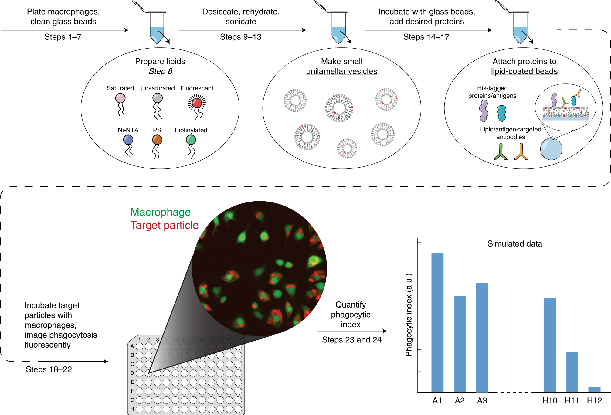 Macrophage Phagocytosis Assay With Reconstituted Target Particles Nature Protocols
 Labeling Of Cd11b Cells By Magnetic Beads And Capture Of Them In A Scientific Diagram
 Development Of Ligand Coated Beads To Measure Macrophage Antimicrobial Activities Tram these cool Biology Of The Cell Wiley Library
1
 Trpv4 Mediates Lpsstimulated Macrophage Phagocytosis Of Igg Coated Scientific Diagram
 Development Of Ligand Coated Beads To Measure Macrophage Antimicrobial Activities Tram these cool Biology Of The Cell Wiley Library
 A Calibration Beads Coated With Either Murine Rat Or Human Igg The Scientific Diagram
 Tcr Activation By Anti Cd3 Coated Beads Impairs T Cell Polarization And Scientific Diagram
Plos One Lilra2 Selectively Modulates Lps Mediated Cytokine Production And Inhibits Phagocytosis By Monocytes
 Isolation Of Tcr Immune Complexes Using Anti Cd3 Cd28 Coated Beads Scientific Diagram

 Bead Based Flow Cytometry For Semi Quantitative Analysis Of Complex Membrane Vesicle Populations Released By Bacteria And Host Cells Sciencedirect
 Magnetic Beads Coated With Gfp Tagged Cbd Proteins Bind And Immobilize Scientific Diagram
 Biomedicines Full Text Exocas 2 Rapid And Pure Isolation Of Exosomes By Anionic Exchange Using Magnetic Beads
 Phagocytosis Of Igg Coated Polystyrene Beads By Macrophages Induces And Requires High Membrane Order Magenau 2011 Traffic Wiley Library
 Cd4 T Cells Isolation Using Antibody Coated Magnetic Beads The Scientific Diagram
 A Method Using Angiotensin Converting Enzyme Immobilized On Magnetic Beads For Inhibitor Screening Sciencedirect
 Magnetic Beads For Protein Antibody Nucleic Acid Purification