Fluorescent Beads Phagocytosis
The Vybrant Phagocytosis Assay Kit provides a way for researchers to observe and quantitate the process of phagocytosis in human polynuclear cells and mouse macrophages by following the internalization of a foreign particlein this case killed E. GP images were pseudo-colored so that yellow to red indicates highly ordered membranes while green represents fluid membranes Figure 1A.
 Development Of A Flow Cytometric Assay To Assess Phagocytosis Of Scientific Diagram
15 Unique, and Fun Fluorescent beads phagocytosis

Potent phagocytic capacity measured by number of beads each cell ingests is shown in both histograms and scatter plots.. GLCM on SIM images revealed a temporal decrease in ASM and IDM Fig. Fluorescent monodisperse latex beads and a computer-centered spectrofluorimeter were used to devise a sensitive new assay for phagocytosis. Representative samples of PBL HKL and SL incubated with fluorescent beads are shown in Figure 5.
Learn More about Flow Cytometry Reference StandardsSection 232. Sandra Casinghino Pfizer Worldwide Research and Development Drug Safety Research and Development Groton CT 06340 USA. These microspheres are packaged as 1 wv suspension in deionized DI water 1mL contains 10mg of fluorescent particles.
Flow cytometric evaluation of phagocytosis of fluorescent latex beads represents a simple rapid nonradioactive index of thyroid function in vitro. In this study we describe a. Specific endocytosis may simply be determined by measurement of the difference in fluorescent bead.
Bangs fluorescent particles have been used in phagocytosis and neural retrograde transport studies and as markers for cell bound antigens. Bead dyes are spectrally matched to common lasers and are highly stable. 6 f as beads were uptaken.
Simplified description of the method and its applications Fluorescent latex beads are easily utilized to quantitate phagocytosis in cells with high endocytic rates such as LM fibroblasts. Another method used injection of fluorescent latex beads into rats followed by removal Address for correspondence. These fluorescent microspheres provide flow cytometry users the means to calibrate laser alignment compensation flow rate dynamic range and other daily adjustments.
The cells were observed and analyzed by fluorescence microscopy sample size 300 cells and flow cytometry sample size 50000 cells to determine the percent phagocytic cells number of beads per phagocytic cell and beads per 100 PMNs. The engulfed fluorescent beads can be detected at the single-cell level. Fluorescent monodisperse latex beads and a computer-centered spectrofluorimeter were used to devise a sensitive new assay for phagocytosis.
However to date no published method quanti-fies particle uptake in real time which is necessary to fully assess the kinetics of phagocytosis. 8772249 PubMed - indexed for MEDLINE Publication Types. Thus was not indicative of phagocytosis.
LM fibroblasts a transformed cell line with a high endocytic rate were exposed to fluoresbrite beads and the following parameters were investigated. Carboxylate-modified polystyrene latex beads fluorescent orange have been used to block phagocytosis by hemocytes in Drosophila. LM fibroblasts a transformed cell line with a high endocytic rate were exposed to fluoresbrite beads and the following parameters were investigated.
In addition the flow cytometric readout provides the advantage of visualizing perturbations in phagocytosis on the population level and when combined with antibody staining of specific cell types within complex populations. It has also been used to assess the uptake of latex beads by antigen presenting cells from porcine respiratory system. To induce phagocytosis IgG-coated polystyrene beads with a diameter of 6067 µm were incubated with Laurdan-labelled macrophages.
Phagocytic activity was stimulated by both cAMP-dependent and cAMP-independent pathways. Beads bacterial and yeast particles labeled with pHrodo red and green were tested for their uptake by THP-1 cells and primary human macrophages by flow cytometry and high-content imaging. 6 d e and increase in entropy Fig.
Coli K-12 strain cells that have been labeled with the fluorescent dye fluorescein. Cells containing beads varied in size morphology and numerous beads ingested as shown by microscopy of Colorrapid stained. Incubation time incubation temperature and beadcell ratio.
After 18 h LPS treatment increased microglia phagocytosis of fluorescent 100 nm beads showing a time-dependent uptake that was not observed in untreated CTRL microglia Fig. Phagocytosis has been widely studied by monitoring the uptake of fluorescent beads or bacteria of 1030 lm in diam-eter or length. In conclusion the bead-based assay for measuring Fcγ-mediated phagocytosis has advantages over assays using intact IE namely fluorescent beads.
We comprehensively evaluated and optimized phagocytosis assays using particles labeled with fluorescent pH-sensitive pHrodo dyes facilitating the specific detection of phagocytosed particles. The ability of FRTL-5 rat thyroid follicular cells to engulf latex beads by phagocytosis was evaluated using flow cytometry and compared to iodide trapping in response to selected growth factors s. Incubation time incubation temperature and beadcell ratio.
Potent phagocytic capacity measured by number of beads each cell ingests is shown in both histograms and scatter plots.
 Phagocytosis Of Latex Beads Notes Counting Of Macrophage Phagocytic Scientific Diagram
 Internalization Of Latex Beads In Phagosomes Phagocytic Assay In Human Scientific Diagram
 Mpl Stimulates Phagocytosis In Microglia And Monocytes Bv 2 Microglia Scientific Diagram
 Microglial Phagocytosis Assay Bio Protocol
 Analysis Of Phagocytosis Inhibition Using Cypher5e Latex Beads A Scientific Diagram
 Phagocytosis Assay Confocal Microscopy Images And Flow Cytometry Scientific Diagram
 Defective Macrophage Phagocytosis Of Bacteria In Copd European Respiratory Society
Latex Beads Covalently Linked To Cps Inhibit The Phagocytic Capacity Of Scientific Diagram
Phagocytosis Of Fluorescent Beads And E Coli Particles A Fluorescence Scientific Diagram
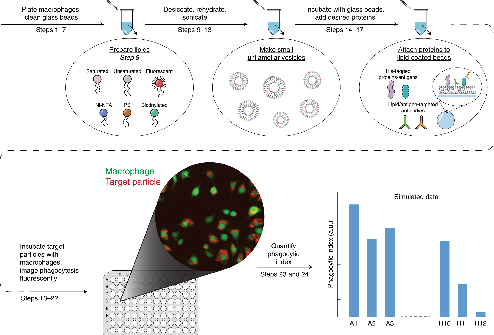 Macrophage Phagocytosis Assay With Reconstituted Target Particles Nature Protocols
 Microparticles Phagocytosis By Dthp 1 Cells Phagocytosis Assay Using Scientific Diagram
 Phagocytosis Of Igg Coated Polystyrene Beads By Macrophages Induces And Requires High Membrane Order Magenau 2011 Traffic Wiley Library
 In Vivo Phagocytosis Of Fluorescent Latex Beads And Fluorescent E Coli Scientific Diagram
 Phagocytosis By Cd4 1 And Cd4 1 2 Myeloid Cells With Fluorescent Scientific Diagram
 Flow Cytometric Analysis Of Phagocytosis Of Latex Beads By Cells Scientific Diagram
 Phagocytosis Of Igg Coated Polystyrene Beads By Macrophages Induces And Requires High Membrane Order Magenau 2011 Traffic Wiley Library
 Trpv4 Mediates Lps Stimulated Macrophage Phagocytosis Of Igg Coated Scientific Diagram
 Hypercapnia Impairs Phagocytosis Of Polystyrene Microspheres And Scientific Diagram
1
 Trpv4 Mediates Lpsstimulated Macrophage Phagocytosis Of Igg Coated Scientific Diagram
 Phagocytosis In Human Macrophages Phagocytosis Of Fluorescent Beads Scientific Diagram
 Latex Beads Phagocytosis By Bm Mscs And Rpe Cells The First Picture In Scientific Diagram
 Phagocytosis Of Cypher5e Labeled Particles A Phagocytosis Of 3 Mm Scientific Diagram
 Protonex Red 600 Latex Bead Conjugate Aat Bioquest