Fluorescent Latex Beads Phagocytosis
LM fibroblasts a transformed cell line with a high endocytic rate were exposed to fluoresbrite beads and the following parameters were investigated. Thus was not indicative of phagocytosis.
Top 15 Best with Fluorescent latex beads phagocytosis
Coli K-12 strain cells that have been labeled with the fluorescent dye fluorescein.. Fluorescent monodisperse latex beads and a computer-centered spectrofluorimeter were used to devise a sensitive new assay for phagocytosis. Fluorescent monodisperse latex beads and a computer-centered spectrofluorimeter were used to devise a sensitive new assay for phagocytosis. Technical advances in the assessment of phagocytosis have allowed rapid advan-.
In addition the flow cytometric readout provides the. Simplified description of the method and its applications Fluorescent latex beads are easily utilized to quantitate phagocytosis in cells with high endocytic rates such as LM fibroblasts. Comparison with iodide trapping as an index of functional activity of thyrocytes in vitro.
Concentration-dependent uptake of beads. If using fluorescent beads. After 1 h of incubation cells were washed and the mean fluorescence intensity was measured by flow cytometry.
After 1 h of incubation uptake of the beads was stopped by incubating the cells on ice and the cells were analyzed by flow cytometry. Readily phagocytose beads even without differentiation into macrophages. Phagocytosis of fluorescent beads by rat thyroid follicular cells FRTL-5.
Phagocytic particle uptake was calculated from data obtained by microscopy and flow cytometry utilizing commercially fluorescent beads of uniform 2 micrometer diameter size. Incubation time incubation temperature and beadcell ratio. Sagartz JE1 Ozaki A Capen CC.
Latex beads carboxylate-modified polystyrene fluorescent yellow-green has been used. If using fluorescent Aβ42. The Vybrant Phagocytosis Assay Kit provides a way for researchers to observe and quantitate the process of phagocytosis in human polynuclear cells and mouse macrophages by following the internalization of a foreign particlein this case killed E.
However although phagocytosis of fluorescent latex beads by phagocytes have been studied for several decades 79 there has not been a reliable quantitative method to measure phagocytic ability of peripheral blood leukocytes for clinical application. Carboxylate-modified polystyrene latex beads fluorescent orange have been used to block phagocytosis by hemocytes in Drosophila. Phagocytosis of the pHrodo green-labeled beads was completely blocked by incubation on ice Figure 1D.
Latex beads were labeled with either pHrodo red or pHrodo green and incubated with THP-1 cells at various concentrations. Incubation time incubation temperature and beadcell ratio. In epidermal growth factor conjugate preparation for indirect immunofluorescence imaging studies for culturing mouse bronchoalveolar macrophages and human monocyte-derived macrophages prior to phagocytosis.
We conducted an experiment to determine the optimum bead concentration to add to cultures and to assess differences between fed. LM fibroblasts a transformed cell line with a high endocytic rate were exposed to fluoresbrite beads and the following parameters were investigated. PMNs collected from a Ficoll-Hypaque density gradient were incubated with the fluorescent beads in human plasma and gently washed in buffer to remove most of the extracellular beads.
It has also been used to assess the uptake of latex beads by antigen presenting cells from porcine respiratory system. Dilute the bead-containing FBS with DMEM to reach the final concentrations for beads and FBS in DMEM of 001 vv and 005 vv respectively. However although phagocytosis of fluorescent latex beads by phagocytes have been studied for several decades 7-9 there has not been a reliable quantitative method to measure phagocytic ability of peripheral blood leukocytes for clinical application.
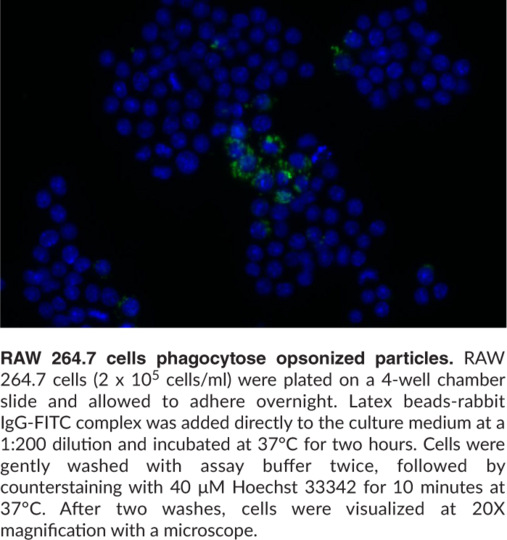
Caymans Phagocytosis Assay Kit IgG FITC employs latex beads coated with fluorescently-labeled rabbit IgG as a probe for the measurement of the phagocytic process in vitro. The ratio of beads to FBS is 15. The ability of FRTL-5 rat thyroid follicular cells to engulf latex beads by phagocytosis was evaluated using flow cytometry and compared to iodide trapping in response to selected growth factors s.
Latex beads labeled with pHrodo red A B or pHrodo green C D were added to THP-1 cells. The engulfed fluorescent beads can be detected at the single-cell level. Specific endocytosis may simply be determined by measurement of the difference in fluorescent bead.
We hypothesized that carboxylatemodified blue fluorescent latex beads could be used to assess rates of feed and bacteria uptake by protozoal cultures. The percentage of phagocytosing macrophages and the number of internalized microspheres per cell was determined from cell size and fluorescence histograms. SUMMARY The phagocytosis of uniform fluorescent latex particles by resident and thiogiycollateelicited macrophages was analysed by flow cytometry.
Another method used injection of fluorescent latex beads into rats followed by removal Address for correspondence. 1Department of Veterinary Biosciences Ohio State University Columbus 43210 USA. Pre-opsonize aqueous green fluorescent latex beads in FBS for 1 h at 37 C.
Sandra Casinghino Pfizer Worldwide Research and Development Drug Safety Research and Development Groton CT 06340 USA. Coli K-12 strain cells that have been labeled with the fluorescent dye fluorescein.
 Phagocytosis Of Latex Beads Notes Counting Of Macrophage Phagocytic Scientific Diagram
 Microglial Phagocytosis Assay Bio Protocol
 Opn Enhances Phagocytosis Of Latex Beads Via The X Integrin Receptor Scientific Diagram
 Phagocytosis By Cd4 1 And Cd4 1 2 Myeloid Cells With Fluorescent Scientific Diagram
Latex Beads Covalently Linked To Cps Inhibit The Phagocytic Capacity Of Scientific Diagram
 Streptococcus Suis Capsular Polysaccharide Inhibits Phagocytosis Through Destabilization Of Lipid Microdomains And Prevents Lactosylceramide Dependent Recognition Infection And Immunity
 Macrophage Reprogramming Influence Of Latex Beads With Various Functional Groups On Macrophage Phenotype And Phagocytic Uptake In Vitro
Plos One Bone Marrow Derived Mesenchymal Stem Cells Maintain The Resting Phenotype Of Microglia And Inhibit Microglial Activation
 Phagocytosis Of Igg Coated Polystyrene Beads By Macrophages Induces And Requires High Membrane Order Magenau 2011 Traffic Wiley Library
 Phagocytosis Of Igg Coated Polystyrene Beads By Macrophages Induces And Requires High Membrane Order Magenau 2011 Traffic Wiley Library
 Pma Activated Thp 1 Cells Allow Investigation Of Phagocytosis Of Scientific Diagram
 Internalization Of Latex Beads In Phagosomes Phagocytic Assay In Human Scientific Diagram
 Phagocytosis Assay Kit Igg Fitc Cayman Chemical
 Protonex Red 600 Latex Bead Conjugate Aat Bioquest
 Pma Activated Thp 1 Cells Allow Investigation Of Phagocytosis Of Scientific Diagram
 Lis1 Regulates Phagocytosis By Localising With Actin In The Phagocytic Cup Biorxiv
 In Vivo Phagocytosis Of Fluorescent Latex Beads And Fluorescent E Coli Scientific Diagram
 Monocyte Subsets Display Different Capacity Of Phagocytosis Which Scientific Diagram
 Gth Enhances Phagocytosis Of Apoptotic Cells Raw 264 7 Cells Were Scientific Diagram
 Figure 2 From Fusion Fission And Secretion During Phagocytosis Semantic Scholar